
If you want to improve your upper body strength or prevent overloading injuries, it is essential to include targeted forearm exercises in your program. The forearm muscles control hand and wrist movements and contribute significantly to the grip thickness, sporting performance and daily function.
From athletes to office workers, anyone can benefit from stronger forearms. Exercises that are aimed at the wrist and forearm muscles can prevent injuries, improve performance and increase general arm development.
overview: What do forearm muscles do?
Your forearm muscles Early movement in your arms, wrists and fingers – and are fundamental for Grip. Your forearm muscles support complex movements such as:
- Turn the palm of the palm up/down
- Bend/expand the wrist and the finger
- Fingers and thumb moved in precision handles
- Stabilization of the hand while gripping or lifting
You are activated daily tasks Like wearing food, writing, lifting weights, using a computer mouse or playing musical instruments.
Key hand joint and forearm movement terms
Stress not about memorizing of all muscles. Remember:
- Flexors (front): Bend your wrist and fingers
- Extensors (back): Stretch the wrist and fingers
- Supination: Turn the forearm so that the palm of your hand goes up.
- Pronation: Turn the forearm so that the palm of your hand flows down.
- Radial deviation (Wrist removal): Tip the wrist to the thumb side.
- Ulnar deviation (Addition wrist): The wrist tends to the small finger side.
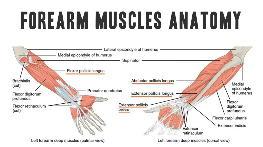
Introduction: anatomy of the forearm muscles
The forearm is the area of the upper extremity between the elbow and wrist. It contains 20 skeletal musclesEveryone that contributes to movements of the elbow, wrist and finger ((1))). These muscles are organized in anterior (flexorter) And Posterior (Extensor Supinator) Subjects, each divided into superficial and deep layers.
Muscle departments of the forearm
1. Front (flexor pronator) subject
Mainly responsible for the flexion of wrist and fingers as well as for Forearm pronation. Every subject contains superficial And deep layersSeparated by bones and a fibrous membrane.


A-) superficial layer (origin near the elbow):
These are the muscles (or structures) that are located on the surface of the body, exactly under the skin and the subcutaneous fat. They are often the most visible or noticeable muscles.
- Pronator Teres: Turns the forearm inside.
- Flexor Carpi Radialis: Bend the wrist and kidnap it (radial deviation).
- Palmaris Longus: Helps with the wrist flexion.
- Flexor Carpi Ulnaris: Bend and add the wrist.
- Flexor Digitorum: Part ((2))).
The Palmaris Longus is missing in about 15% of the population. Its absence does not affect the grip or wrist function ((3))).
C-) Deep layer:
These are the muscles (or structures) the farthest from the surface, typically directly on or very close to the bone.
- Flexor Digitorum deep: Bend the distal finger joints of the finger 2–5.
- Flexor thumb longus: Bend your thumb.
- Pronator Square: Turns the forearm inside (pronation).
2. Posterior (Extensor Supinator)
These muscles extend The wrist and fingers help with Supination (Palm-up rotation) and help to stabilize the wrist. Most come from the Lateral epicondyle of humerus.


A-) superficial layer:
- Brachioradialis: Bend the elbow, helps with rotation.
- Extensor Digitorum Longus & Kurz: Expand and kidnap the wrist.
- Extensor Digitorum: Expand the fingers.
- Extensor Digitorum: Expand the little finger.
- Extensor Carpi Ulnaris: Advanced and adds to the wrist.
- Anconeus: Helps to expand the forearm on the elbow.
The Extensor Digitorum split into three slips: one is on Middle Phalanxand two merge on Distal phalanx.
B-) Teeme layer:
- Supinator: Turns the forearm so that the palm of your hand drives up (supination).
- Extensor Digitorum longus: Expand your thumb.
- Longus kidnapper: Moves the thumb away from the palm.
- Extensor: Expand the index finger.
Common wrist and forearm conditions
The wrist and the forearm consist of a complex network of bones, muscles, tendons, ligaments and nerves, enable fine motor control and strong grip. These structures are susceptible to overuse, trauma and attitude, especially for athletes, desk workers and manual workers.
Understanding the most common diseases that influence this region is of essential importance for early intervention, adequate treatment and effective rehabilitation.
| Condition | Caused | Key symptoms | Treatment |
|---|---|---|---|
| Carpal tunnel syndrome | Median nerve -nerve -compression on the wrist | Dust in the thumb/index/middle finger, night pain | Shelf, nerve slides, surgery, if serious |
| Tennis -Ellbogen (lateral epicondylitis) | Overuse of wrist extensions | Pain on the outer elbow, weak grip | Quiet, eccentric exercises, forearm belt |
| Golfers Ellbogen (media epicondylitis) | Overuse of wrist flexors | Pain on the inner elbow, sore with wrist flexion | Quiet, stretching, eccentric bumping training |
| From Quervain’s tenosynovitis | Inflammation of APL/EPB tendons | Pain near the thumb base, worse with a thumb movement | Thumb rail, NSAIDS, corticosteroid injection |
| Wristspin | Band pollution or tear | Swelling, bruising, limited Rome | Rice, tension, gradual rehabilitation |
| Distal radius fracture (Colles’) | Fall out | Wrist deformity, pain, swelling | Castle or surgical fixation |
| Ulnar nerve connection (Guyons Channel) | Compression of the ulnar nerve on the wrist | Dust in fink/ring fingers, handle weakness | Upholstery, nerve slides, surgery if necessary |
| Flexor tendinitis | Overuse of wrist/finger flexors | Pain during the wrist/finger flexion | Rest, NSAIDS, stretching |
This is how they strengthen their forearms
Try this Forearm duration exercises at home or in the gym:
- Hand gripper or tennis ball: Improvement of squeezing strength.
- Forearms, curls curl: Use light weights to curl with your wrists.
- Wrist roll: A cheap and effective way to strengthen the forearm muscles.
- Fidelity: Snap yourself dumbbells and go and concentrate on the grip.
- Pobilizer/pull -ups: Great for forearm, handle and biceps.
- Reverse curls: With a rollover, curl dumbbells or a bar.
- HAMMER curl: Great for brachioradialadial and wrist stabilizers.
- Kreuzleben, rows, kettlebell fluctuations: Build up overall pulling strength.


Checkout Popeyes forearm training For stronger forearms
Forearm muscle stripes
Relieve tension and prevent them with these injuries:
- Wrist rotations: Make fists, turn the wrists in both directions.
- Forearm: Arm straight, pine up, pull it down gently.
- Route of the forearm: Arm straight, hand down, pull your hand back carefully.


The strengthening and stretching of your forearms can promote a better function and prevent an injury.
- Do not neglect them in your training.
- Train both Beuger and extensors.
- Treat injuries early.
- Keep them mobile and resilient with routes.
Diploma
The forearm is a complex region that consists of 20 muscles that enable precisely and powerful movements of the wrist, hand and digit. Understanding the layered anatomy, functions and variations is essential for medical specialists, fitness specialists and everyone who is interested in biomechanics for human people. The detection of frequent anatomical variants and clinical implications improves diagnostic and therapeutic accuracy.
References
- Brittney Mitchell, Lacey Whited. Anatomy, shoulder and upper extremity, forearm muscles
- Lauren Okafor, Matthew A. Varacallo. Anatomy, shoulder and upper extremity, hand flexor digitorum Oberficialis muscle
- Prevalence of the Palmaris Longus muscle and its relationship with the handle and pinch of strength: a study in a Turkish pediatric population
- Moore Kl, Dalley Af, Agur am. Clinically oriented anatomy. 7th edition Lippincott Williams & Wilkins.
- Standring S, Ed. Gray’s Anatomy: The anatomical basis of clinical practice. 41. Ed.
- Saladin KS. Anatomy & physiology: the unity of shape and function. 9. Ed.
- Nice FH. Atlas of human anatomy. 7. Ed.
- Jacobson MD, Raab R., Fazeli BM, et al. Anatomical variations of the forearm muscles and their clinical importance. Clin Anat. 2001.
- Roy J. variants of the palaris longus muscle. J Hand Surg. 1995.
- Sunderland S. Nerve and nerve injuries. Volume 1 & 2nd 1978.
- Hirasawa y, et al. Studies on the distribution of the superficial branch of the radial nerve. J Hand Surg am. 1987.
- Thompson NW, Mockford BJ, Cran Gw. A lack of the Palmaris Longus muscle. Ulster Med J. 2001.
- Roy TS, et al. Variation of the origin of the Flexor Pollici Longus. Clin Anat. 2002.
- Oh SJ, et al. The Martin Gruber anastomosis in humans. Muscles. 1976.